

Magnetic Resonance Imaging (MRI), CT and X-ray exams can sound similar to patients who are preparing for an appointment, but each test works differently. The machines look different, the scan times vary, and the reasons a doctor orders one test instead of another are not always obvious. Kasey McKillip, a seasoned MRI technologist with over a decade of hands-on experience in medical imaging, recognizes that patients often feel more comfortable when they understand the basic purpose of the exam before they arrive. Kasey McKillip explains that understanding the differences between these exams can make appointments feel less confusing for patients.
The goal is not to memorize medical terms. It is to have a clearer idea of what the test may involve, why certain safety questions matter, and what kind of information the scan may help provide. A patient who understands the difference between MRI, CT and X-ray imaging may feel less caught off guard when preparing for the appointment.
Why Doctors Use Different Imaging Tests
Doctors order imaging exams when they need more information about what may be happening inside the body. Symptoms such as pain, swelling, weakness, injury or shortness of breath may point to several possible causes. Imaging can help the care team look more closely at bones, organs, soft tissue, blood vessels or other areas that cannot be fully assessed from the outside.
No single imaging test is best for every situation. MRI, CT, and X-ray imaging each have different strengths. The test a doctor chooses depends on the body part involved, the medical question being asked, the urgency of the situation, and the type of detail needed for review.
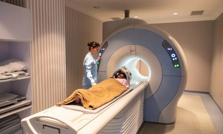
What MRI Imaging Is Often Used For
MRI uses a strong magnetic field and radio waves to create detailed images of the body. It does not use ionizing radiation. MRI is often used when doctors need a closer look at soft tissues, the brain, spine, joints, muscles, ligaments, or certain organs. The scan can take longer than some other imaging exams and usually requires the patient to stay very still.
For patients, the MRI experience can feel more involved because of the loud sounds, enclosed space and careful safety screening. The technologist may ask about implants, metal fragments, pacemakers, surgical clips, or medication patches. These questions are part of the process because the magnet is powerful and must be treated with care.
What CT Imaging Is Often Used For
CT stands for computed tomography. A CT scan uses X-rays and computer processing to create cross-sectional images of the body. The scan is often faster than an MRI and may be used in emergency settings, injury evaluations, or when doctors need a quick look at areas such as the head, chest, abdomen, or bones.
Some CT exams use contrast material, which may be given through an IV or taken by mouth, depending on the exam. Patients may be asked about kidney problems, allergies or prior contrast reactions. CT scans do involve ionizing radiation, so the care team considers the medical reason for the test before ordering it.
What X-Ray Imaging Is Often Used For
X-ray imaging is one of the most familiar types of medical imaging. It is often used to look at bones, joints, chest, or certain injuries. Many patients have had an X-ray after a fall, sports injury or illness affecting the lungs. The exam is usually quick and may involve standing, sitting, or lying in a specific position.
X-rays use a small amount of ionizing radiation to create images. The technologist may ask the patient to hold still or hold their breath for a moment, depending on the body part being imaged. While the test itself is often brief, careful positioning still matters because the image needs to show the correct area.
How the Patient Experience Can Feel Different
The patient experience can vary quite a bit across MRI, CT, and X-ray appointments. An X-ray may be finished quickly, while a CT scan may take a little longer, depending on preparation and contrast. An MRI often takes more time because it captures many detailed image sequences and requires careful positioning.
Kasey McKillip’s patient-centered approach reflects the importance of explaining these differences before the scan begins. A patient may feel less nervous when they know why one exam takes longer, why another uses contrast, or why certain questions are asked. Clear information can make the process feel more organized rather than confusing.
Why Safety Screening Is Different for Each Test
Safety screening changes depending on the type of imaging exam. For MRI, metal and implanted devices are a major concern because of the strong magnetic field. Patients may need to remove jewelry, watches, hearing aids, hairpins, belts, phones and clothing with metal parts before entering the scan room.
For CT and X-ray exams, radiation exposure and pregnancy status may be part of the safety conversation. For CT exams with contrast, kidney function and prior contrast reactions may also matter. These questions are not meant to alarm patients. They help the imaging team choose the safest way to complete the exam based on the patient’s situation.
How Results Are Reviewed
After an imaging exam, patients may want immediate answers. That reaction is understandable, especially when the test is connected to pain, illness, or uncertainty. The technologist usually cannot interpret the images or provide a diagnosis during the appointment.
A radiologist reviews the images and prepares a report for the provider who ordered the exam. That provider discusses the results with the patient and explains what they may mean in context. The timing can vary by facility and situation, so patients should ask when results may be available and who they should contact with questions.
Understanding the Test Can Ease Uncertainty
MRI, CT, and X-ray imaging each play a different role in patient care. MRI is often used for detailed soft tissue views. CT can provide fast cross-sectional images. X-ray is commonly used for bones, joints and chest imaging. Each test has a purpose, and the choice depends on what the doctor needs to understand.
The most helpful step is to ask practical questions before the appointment. What test is being done? Is contrast expected? How long might it take? Are there safety concerns to mention? Clear answers can make the imaging process feel less unfamiliar and help patients arrive with a better sense of what to expect.


